Quad Screen Blood test for Down, Trisomy 18, and neural tube defects
Also known as the “4-Marker Screen,” this is a commonly offered screening test for some genetic problems.
This is offered
between 15 and 22 weeks and measures four substances: alpha-fetoprotein
(AFP), human chorionic gonadoptropin (hCG), estriol, and inhibin-A. If
the levels are low or high, this can indicate an increased risk for
genetic problems and you will be offered further testing.
Should I have a scan? Is ultrasound safe in pregnancy?
Ultrasound scans in pregnancy, first introduced 40 years ago, have become a routine part of prenatal care.
Most research indicates that they are a safe way to view the baby, even when extra scans are needed for medical reasons.
Suggested
links between additional scans and growth problems and dyslexia are
tentative since babies scanned more often are more likely to have
problems linked to other factors.
Recommendations are that scans are performed only for clinical reasons and the number done is kept to a minimum.
3D and 4D ultrasounds
Many companies now
offer special scans that reveal your baby in three dimensions or moving
on film or video. These 26–32 week scans can be quite expensive and are
done for curiosity value and not for medical reasons. The quality of the
pictures is usually amazing and parents are sometimes able to spot
genetic similarities between themselves and their baby. However, the
scan is often lengthy, which means the baby is exposed to ultrasound for
longer than is normal. Also, if the baby is in the wrong position, it
may be difficult to get a clear picture. The position of the placenta,
the amount of amniotic fluid, and the size of the mother can also affect
the quality of the pictures obtained.
Moving pictures
These detailed
scans offer incredible clarity, often revealing family resemblances and
sometimes enabling parents to see their baby moving around, perhaps
sucking its thumb, rubbing its eyes, or yawning.
NOTE
Despite the concerns that often accompany screening tests, try not to let these overshadow the joy of being pregnant
NOTE
Knowing that you are receiving such thorough prenatal care throughout your pregnancy can be deeply reassuring
Nuchal translucency and dating scans Ultrasound examinations
An ultrasound can be
performed in the first trimester if menstrual dates are uncertain but in
some locations it may be requested for all patients. Because the nuchal
translucency scan has a small window of time in which it can be
performed, 11–14 weeks in most centers, a dating ultrasound may be done
prior to first trimester genetic screening.
| Q: |
What does the dating ultrasound look for?
|
| A: |
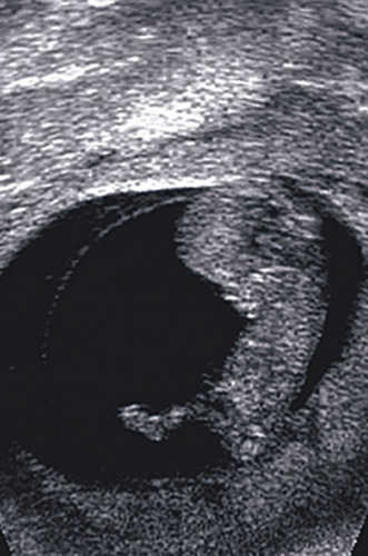
The distance is measured from the top of the baby's head to its
bottom (crown—rump measurement), and the diameter of the head is
recorded, known as the biparietal diameter—the distance between the
parietal bones either side of the head.
|
| Q: |
How is the nuchal translucency scan done?
|
| A: |
The nuchal translucency test is performed through the
application of ultrasound targeted at the back of the baby's neck. The
technician will measure the thickness of any fluid collected behind the
baby's neck. In general, a thickness of 2–3 mm or less can be considered
normal. In the first trimester, there is an association between the
size of fluid collection at the back of the fetal neck and risk of Down
syndrome. If early testing is declined or if prenatal care starts too
late, the quad screen may be the best option, given between 15–22 weeks.
|
| Q: |
Is it reliable?
|
| A: |
The nuchal translucency scan is considered to be 80 percent
accurate, which means there is a 20 percent (1 in 5) chance of it being
inaccurate. If you're offered a blood test (PAPP-A) with the scan, it
becomes 85 to 90 percent accurate. When the nasal bone is also measured,
the accuracy rises to 95 percent. Your health-care provider should
provide you with information the reliability of these screening tests.
|
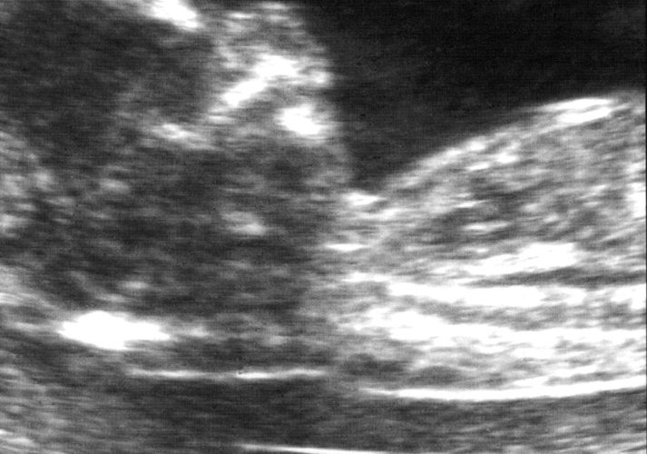
This fetus has a small amount of fluid behind the neck (seen as
the black area at the back of the neck), which suggests a low risk of
Down.

At 12 weeks, the fetus has a clearly defined profile with a prominent nasal bone.

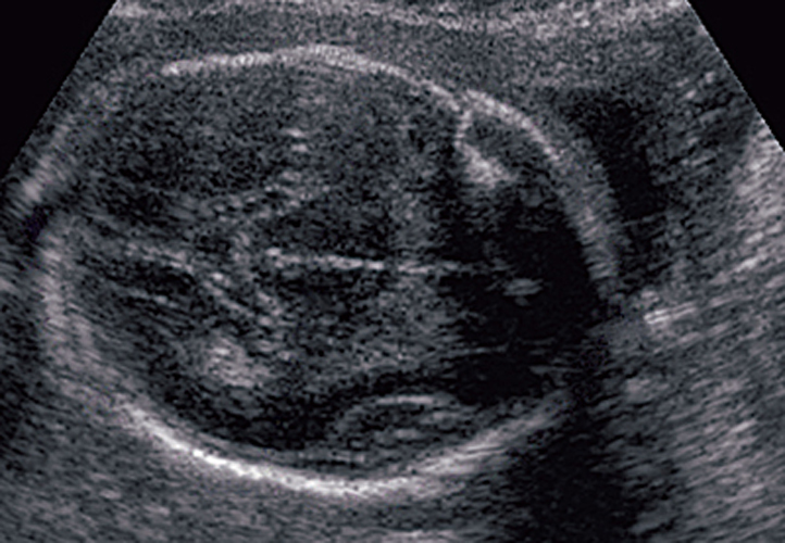
One of the measurements taken to plot fetal growth is the
biparietal head diameter: the distance between the two head bones.
