Your 20-week Scan
This second-trimester
ultrasound, typically done between 18–22 weeks, looks in detail at how
your baby’s major organs and body systems have developed, as well as
checking the placenta and the volume of the amniotic fluid.
What the scan reveals
By 20 weeks, your
baby’s organs and body systems are well developed and can be seen
clearly on an ultrasound scan. The sonographer performing the scan will
look closely at how your baby’s major organs and body systems have
formed and whether there are any indications of a problem (see Your baby’s checkups). In the majority of cases, the scan will reassure women that their baby is developing normally.
If your baby is
found to have a problem, the sonographer will refer you to a maternal
medicine expert who will confirm the findings and offer follow-up scans
throughout the rest of your pregnancy. He or she will also talk to a
pediatrician to ensure that they have enough information to take care of
your baby at birth.
A picture of your 20-week scan shows the incredible detail visible on an ultrasound at this stage of your pregnancy.

Your due date
If you had a scan in the
first trimester, your dates are unlikely to be changed after a second
trimester scan. This is because dating is most accurate in early
pregnancy when all babies essentially grow at the same rate. Later on,
individual differences in growth start to appear, making it harder to
tell if your baby is just big for dates or if you are further along than
thought.
However, if you
didn’t have a first trimester scan, the sonographer may change your
dates if your baby is 10–14 days smaller or larger than expected. If
your dates have been confirmed by a first trimester scan, a substantial
lag may be a sign of a growth problem in your baby , although this is rare.
Measuring your baby
Because your whole baby
no longer fits on the screen, the crown–rump length won’t be measured.
Instead, your baby’s size will be calculated by combining a series of
measurements in a mathematical formula. The sonographer will measure the
width (biparietal diameter) and circumference of your baby’s head, the
circumference of your baby’s tummy (abdominal circumference), and the
length of your baby’s upper leg bone (femur length). These measurements
help estimate the size of your baby and check that this is within the
normal range for this stage of pregnancy.
Placenta and amniotic fluid
The placenta will be
examined to ensure that it appears normal and isn’t blocking the baby’s
exit route (the cervix). It’s quite common in early pregnancy for the
placenta to be low-lying and to cover the cervix, but in 95 percent of
cases, the placenta moves up and out of the way by the third trimester
as the uterus grows.
If your placenta is low
lying, your doctor will arrange a follow-up scan later in pregnancy to
check that the placenta has moved up out of the way. If a later scan
shows that it has failed to move up, a condition known as placenta
previa , you will be monitored until the birth.
The amniotic fluid is
assessed to check that there isn’t too little or too much. If there is
too much fluid, it may be possible to drain some using amniocentesis to decrease the risk of later complications such as premature labor. Too little fluid can indicate a problem with fetal growth or your baby’s renal tract and your baby may need to be monitored.
Markers for Down syndrome
Although ultrasound at this
stage is not a reliable way to detect Down syndrome, it can pick up
certain signs known as “soft markers” that may suggest that your baby
has an increased risk of Down syndrome. However, many markers are very
common and are not usually a cause for alarm unless you already have an
increased risk for Down syndrome, for example, because you are over 35
years of age, or were given a high risk from a previous screening test.
Common markers include a bright spot in your baby’s heart, seen in about
1–2 percent of normal babies; extra fluid in your baby’s kidneys; short
leg or arm bones; thickened neck skin (nuchal fold); and bright
(echogenic) fetal intestines.
Some specific
abnormalities are linked with a far higher risk of Down syndrome or
other chromosomal abnormalities. These include certain kinds of heart
defects and other major malformations, such as abnormalities in the
spleen.
What happens next
If the sonographer detects soft markers, he or she will discuss the findings and may suggest a diagnostic test,
especially if you already have an increased risk of Down syndrome.
However, an earlier all-clear from a diagnostic test means that you can
be confident your baby’s chromosomes are normal. If an obvious
abnormality is seen, and you haven’t had a diagnostic test, this will
probably be strongly advised. Having information from the scan will help
you decide whether you want to proceed with a test. Whatever your
decision, any associated abnormalities will be monitored throughout your
pregnancy.
Your baby’s checkups
What the ultrasound looks at
During this scan,
your baby’s organs are examined in detail and therefore it can take a
little longer to perform than previous scans. For most, the scan
provides reassurance that their baby is developing normally. The
following areas will be checked.
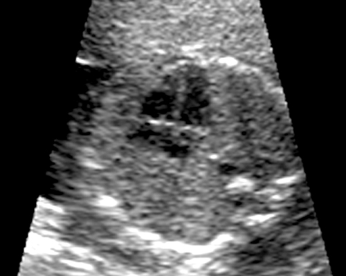
The brain, including the fluid-filled spaces inside the brain and the shape of the back of your baby’s brain (cerebellum).
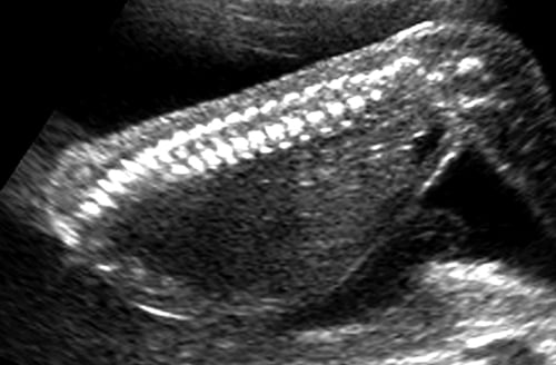
The spine, to check for spina bifida or other problems.
The upper lip, to check for cleft lip.
The heart, to rule out any major heart malformations. The heartbeat will be checked too.
The stomach and diaphragm.
The kidneys and bladder, making sure that both kidneys are present and there are no blockages or malformations.
The abdominal wall, to look for a defect, known as gastroschisis.
Your baby’s limbs, to make sure there are no hand or feet malformations, such as club foot.
The umbilical cord, to check that this has a normal number of blood vessels.
How the scan is interpreted
During your ultrasound, a picture of your baby is produced
when high-frequency sound waves bounce off your baby and translate into
an image on screen. In this image, solid matter, such as bones, are
white, while softer tissue appears gray. Areas that contain fluid, such
as blood vessels or the stomach, as well as the amniotic fluid, do not
respond to the sound waves and therefore appear as black areas on the
scan. The sonographer will study these details to assess how your baby
is developing in the uterus.
The skull is well developed by now, and features such as the ears are clearly seen.
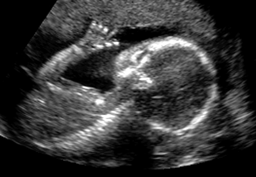
The four chambers of the heart can be identified and certain defects spotted.
The leg bones are measured to assess how your baby is growing.
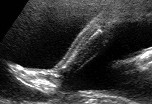
Each vertebra in your baby’s spine will be counted to check for spina bifida.
Boy or girl?
Unless your baby is curled up or the legs
are closed tight, the sonographer will have a pretty good idea of
whether you’re going to have a boy or a girl. If you don’t want to know
the sex, tell the sonographer at the start of the scan. If you do,
consider asking the sonographer to point out the baby’s genitalia
onscreen. If you are told which sex your baby is, resist the urge to go
out and spend money on all-blue or all-pink decor since sonographers
have been known to get it wrong!