Understand when and why your
ultrasounds are done
Diagnosing Down syndrome
A definite diagnosis of Down syndrome can
currently only be made by invasive testing, commonly by amniocentesis or
chorionic villus sampling (CVS). Down syndrome occurs when the baby receives an
extra chromosome 21 (trisomy 21). It is one of the most common serious
abnormalities that occur and is associated with severe mental retardation. It
can occur at any age but is more common in older women.

A
definite diagnosis of Down syndrome can currently only be made by invasive
testing, commonly by amniocentesis or chorionic villus sampling (CVS).
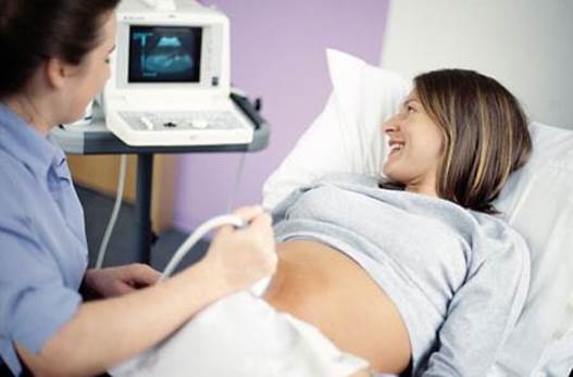
One thing that you can be sure of during
your 40 weeks of pregnancy is that you will be going for numerous doctors’
appointments, and many of these will include ultrasound scans. The number of
scans you will have will depend on whether you are using a private hospital or
a government one, and if you are having a high-risk or straightforward
pregnancy. While seeing your little one moving around your womb is an amazing
feeling, most sonar scans are for more than just getting a baby picture.
Gynaecologist and obstetrician Dr. Serilla Moodley breaks down what you can
expect at your scans, and what they are really there for.
12- and 20-week scans
The two most important scans are the
12-week and 20-week scans. “The first trimester scan can be done between 12
weeks and 13 weeks, six days. We are quite specific about that time frame. In
order for the measurements to be accurate the scans must be done at that time.
We look at many features on the fetus when we do this scan, the most important
being the nuchal translucency. This is a swelling at the back of the fetus’
neck which is a marker for Down syndrome, cardiac abnormalities, pre-eclampsia,
and skeletal abnormalities. The value of the unchallenged translucency test is
that on its own it can predict 70 percent of abnormalities, and when combined
with a blood test they can predict 97 percent.”
While this 12-week scan is commonly
referred to as the nuchal translucency scan, it is also used to examine other
things. “We also look at other features on the fetus such as the facial angle,
nasal bone, and the presence of a bladder... We do cervical length and uterine
artery dopers (which measures the blood flow to the uterus and can be used to
predict pre-eclampsia),” Dr. Moodley says.
The second screening test that is vital for
you to have, whether you are at a private or government hospital, is the
20-week scan. “This can be done between 18 and 24 weeks. It is an anatomy scan
of the fetus and is used to identify any structural abnormalities such as any
heart valve defects, cleft lip/palate, and spinal problems. We also look at
soft markers for chromosomal abnormalities,” Dr. Moodley explains. “A soft
marker is something unusual in a fetus, which in itself is not a problem, but
when there are lots of soft markers present it can be used to predict
chromosomal problems.”

While
this 12-week scan is commonly referred to as the nuchal translucency scan, it
is also used to examine other things
If an abnormality of some kind is detected,
it is important to know that these tests are all only screening tests and
cannot definitively diagnose a problem. If the screening tests come back
abnormal, then you will be offered diagnostic tests such as a chorionic villus
sampling or amniocentesis to shed some more light on the problem, if there is
one.
Overdue scan
A final important scan that you may be
ordered to have is an overdue scan. If you are still pregnant past your due
date, this scan will look at the amniotic fluid volumes, placenta (for
calcifications), growth and fetal weight, and the blood flow in the umbilical
cord. If your caregiver is concerned at all, they will send you for a
non-stress test (NST) which measures whether your baby is under any stress in
the womb. In between your vital scans and the ones that you may choose to have,
you get your routine scans. If you are going through your pregnancy in the
government sector, you may only have these scans if your doctor or midwife
feels that there are any complications, or if your pregnancy has been diagnosed
as high-risk for any reason. Dr. Moodley explains that these scans are done to
follow up the growth of the fetus, to look at amniotic fluid volumes and
monitor placental condition. Caregivers can use these scans to identify growth
restriction in the fetus (IUGR), especially in conditions like pre-eclampsia,
or macrosomia in diabetics, or to monitor the condition of placenta praevia.
Just for fun
There are a few scans that are not entirely
necessary, but are optional to you. The 16-week scan is mostly carried out to
identify the sex of the baby (although a really experienced fetal medicine
sonographer will be able to do this at the 12 week scan). The 3D or 4D scans
are a nice-to-have keepsake in pregnancy, as many features, especially facial
features, can be identified. In proper hands they can be used to identify or
confirm structural or chromosomal abnormalities. But mostly, the 4D scans are
not diagnostic, and you may choose to get one done to have the images or DVD as
a memento.
There
are a few scans that are not entirely necessary, but are optional to you.
For your information
If your 12-week or 20-week scans indicate
any abnormalities, you will have the option of doing either an amniocentesis or
chorionic villus sampling for further diagnostic investigation.
An amniocentesis is done at around 16 weeks.
A needle is passed through the mom’s abdomen, through the uterine wall and into
the fluid that surrounds the baby. A small sample of this fluid is removed.
A chorionic villus sampling is usually done
at around 11 weeks. A needle is passed through the uterine wall into the
placenta and a small sample of the placenta is removed.
In both cases, the sample is sent to the
genetic lab where fetal cells are cultured and the chromosomes are analyzed.